EMERGENCY NUMBER
91-9040092702

Plot-659, Tankapani Rd, Rajarani Colony, BJB Nagar, Bhubaneswar, Odisha 751014

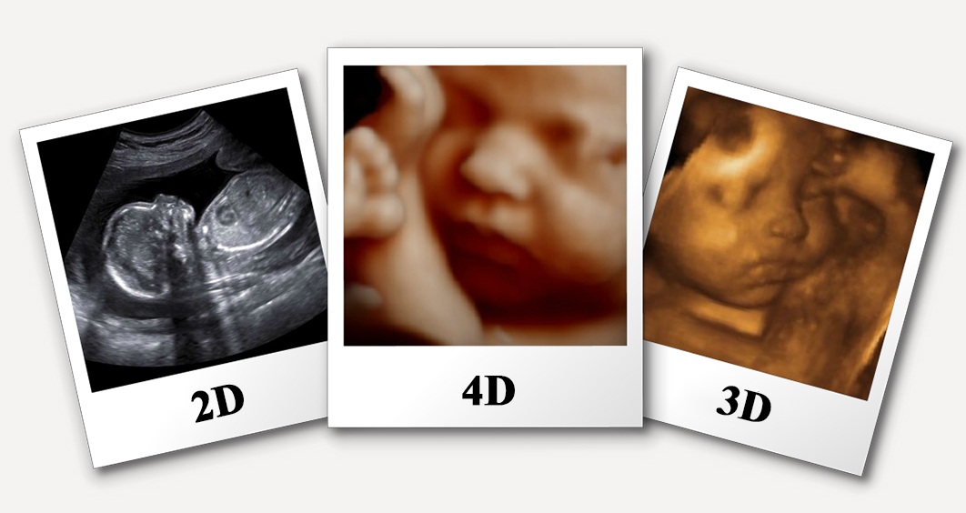
Four-dimensional (4-D) ultrasound is 3-D ultrasound in motion.
Color Doppler ultrasound is a special ultrasound technique that
evaluates blood
flow through a blood vessel, including the body's
major arteries and veins in the abdomen, arms, legs and neck.
Ultrasound is a high-frequency sound that you cannot hear but it can be emitted and detected by special machines.
It travels freely through fluid and soft tissues and is a safe and painless scan.
Our procedures at ShreeUsha Diagnostics conform to world-class standards and PNDT guidelines.

An Ultrasound (solography) is a Test procedure in which high-frequency sound waves (sonar and radio technology) are used to create ‘pictures’ of the inside of the body.
Apart from being safe, an Ultrasound is also a painless and generally affordable process – making it popular with both patients and the medical fraternity.
Ultrasound images are engineered in ‘real-time’, and let us see both the structure of our internal organs as well as the function (that is, the movement) of our blood vessels.
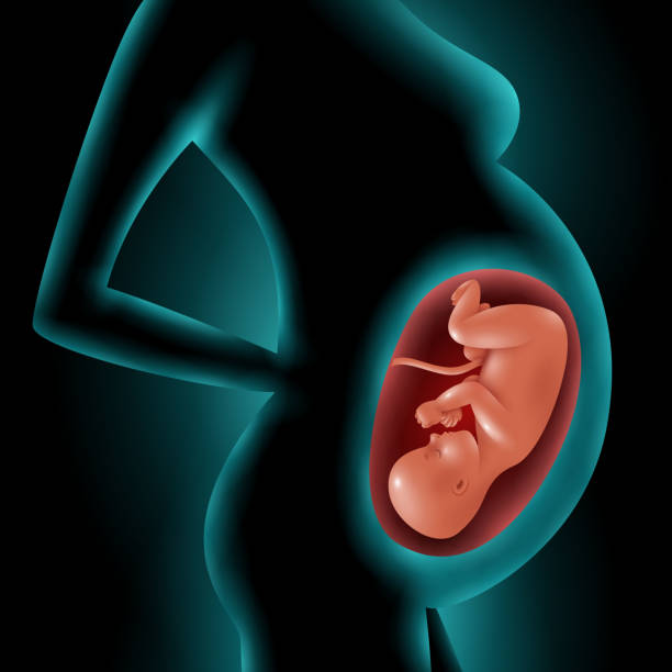
While it has various kinds of medical uses and health applications, an Ultrasound is perhaps the most popular and preferred Test used to monitor the development of the fetus in pregnant women.
An Ultrasound Test is carried out by a doctor or a Radiologist who has been specially trained for the purpose.
An Ultrasound Test usually takes between 15 minutes to an hour, and normally doesn’t require any special preparation.
For an Ultrasound a bladder that is full of water is able to generate better pictures or images of the uterus and other organs (since air is a bad conductor of ultrasound and makes the scan a failure).
In the case of an External Ultrasound, the Transducer is placed over the part of the body (for example, the heart or the abdomen) that needs to be studied and has been administered a lubricating gel before-hand for this purpose.
This process is usually free of any kind of pain or discomfort.
In the case of an Internal Ultrasound where the ovaries or urinary organs need to be studied in greater detail, the Transducer is inserted into the rectum (in the case of males) or the vagina (in the case of females). These studies can be routinely performed in approved outpatient clinics.
The term ‘Ultrasound’ literally means sounds belonging to a ‘frequency’ that humans are not able to ‘detect’ or hear.
For most diagnostic applications, this frequency range lies between 2 and 18 megahertz (MHz).
Higher frequencies can generate better Ultrasound image quality but are susceptible to getting absorbed (and therefore, ‘diminished in power’) by the skin and other soft tissues.
Lower frequencies can penetrate deeper past the layers of skin & tissues but produce imagesmof a relatively lower resolution and quality.
During the process, the Radiologist will hold a wand-like device called the Transducer in his or her hand. The transducer is placed on the patient’s body externally.
In some cases, insertion may be done via the rectum (for males) and vagina (for females) and the Transducer in such situations is ‘placed internally’.
Be it external or internal, sound waves are emitted by the machine which travels through the skin and tissue till it reaches a surface that is too dense for it to penetrate further.
The sound waves are then reflected back (similar to the act of light reflecting off a mirror or, say, an echo of our own voice) – resulting in an image.
This image or picture is also sometimes referred to as a Sonogram. Different shades of grey indicate different densities of the reflecting surface inside the body, which can be an organ or an internal structure.
Despite being different technically, the terms Ultrasound and sonogram are sometimes used interchangeably.
| WHOLE ABDOMEN | OVERNIGHT FASTING / 4-5 HOURS FASTING, FULL BLADDER |
| UPPER ABDOMEN | OVERNIGHT FASTING / 4-5 HOURS FASTING |
| LOWER ABDOMEN / PELVIS | FULL BLADDER, NO FASTING REQUIRED FOR MARRIED PATIENT ADVISE TVS |
| TVS | NO PREPARATION REQUIRED,EMPTY BLADDER |
| USG EARLY PREGANCY / BLEEDING PV | ADVISE TVS |
| USG NT/NB / LEVEL II/ COLOR DOPPLER PREGNANCY | NO PREPARATION |
| USG KUB | FULL BLADDER, NO FASTING REQUIRED |
| USG FOR STONES | |
| FOR GALL BLADDER STONES | 4-6 HOUR FASTING |
| FOR KIDNEY STONES | FULL BLADDER |
| RENAL DOPPLER | 4 HOURS FASTING preferred |
| ABDOMEN DOPPLER | 4-5 HOURS FASTING |
| LOWER LIMB DOPPLER / UPPER LIMB DOPPLER | NO PREPARATION |
| USG SOFT TISSUE / SOFT TISSUE DOPPLER | NO PREPARATION |
| USG SCROTUM | NO PREPARATION |
| USG BREAST | NO PREPARATION |
| USG CHEST | NO PREPARATION |
| WHOLE ABDOMEN | To see for- Liver, kidney, gall bladder, Pancreas, Intestines, infection, stones
|
||||||
| UPPER ABDOMEN | To see for – Liver, kidneys, pancreas, intestines, Stones | ||||||
| LOWER ABDOMEN / PELVIS/TVS | Usually done in females
|
||||||
| PVR | Post void residual urine | ||||||
| KUB | To see for - kidneys ureter, bladder
|
||||||
| Early Pregnancy / TVS for pregnancy | To see site of pregnancy – Uterus, Tubes , Ectopic pregnancy , Gestational sac , Heart beat , Development of fetus , Calculate weeks of pregnancy , Associated fibroid / cyst/ Infection | ||||||
| NT/NB SCAN (LEVEL I) <11-14 week> can be done till 16 weeks> Level I scan, NT scan, Genetic sonogram, Early anomaly scan | Nuchal Translucency / Nasal Bone
To see for:
Diagnosis of Down’s syndrome, Genetic defect, Brain development Our SPECIALITY: I.T., FM ANGLE, BLOOD FLOW TO BABY, EARLY ANOMALY DETECTION, 4D/5D BABY |
||||||
| LEVEL II SCAN <17-24 WEEK>can be done till 28 weeks TIFFA scan, Anomaly scan, 18-22-week scan, Genetic scan, congenital scan | To see for: , Detailed Scan of foetus , Baby face, heart organs, hands, feet Our SPECIALITY: , BLOOD FLOW TO FETUS , IMMERSIVE 4D/ 5D IMAGES | ||||||
| FETAL ECHO <20-28 WEEKS> | To see for: , Fetal Heart , Any congenital Heart defect | ||||||
| BPP / BPS Biophysical Profile, Biophysical Score, Manning’s Score | To see for: , Blood flow to baby , All blood vessels from mother to baby , Growth , Movement. , Fluid level. , Cord around neck. , Placental position. Our SPECIALITY: , 4D/5D Baby – highest resolution available | ||||||
| COLOR DOPPLER | Blood flow through blood vessels | ||||||
| Color Doppler carotid (neck) | To see for: , Blood flow through neck vessels carotid arteries , Carotid arteries are the main source of blood flow to the Brain | ||||||
| Abdomen Doppler | To see for: , Blood supply to liver – cirrhosis, Liver failure, chronic alcoholic , Blood supply to intestines | ||||||
| Renal Doppler | To see for: , Blood supply to kidney , Chronic kidney disease , Hypertension in young , Sometimes pregnancy induced hypertension | ||||||
| Lower limb Doppler Arterial | To see for: , Blood supply heart to foot , Blockage , Narrowing of arteries , Reduced blood flow | ||||||
| Lower limb Doppler venous | To see for: , Blood supply foot to heart , Blockage , Varicose veins | ||||||
| Pelvic Doppler / 3D pelvis Doppler | To see for: , Blood supply to uterus / ovaries , Cysts categorisation | ||||||
| Scrotum Doppler | To see for: , Infertility , Torsion (twisted testis) , Blood supply to scrotum , Infection / mass | ||||||
| 3D TVS/ 4D TVS/ 5D TVS | Better Resolution / Clarity
Better Diagnosis At most places TVS / LA is done by 2D Probe due to which uterine anomalies are easily missed but here it is picked. |
||||||
| Why 4D / 5D Scan | , Transducer Capacity (1-13 MHz) , Higher the Frequency range better the resolution , 1 – 3 MHz Deep Penetration , Higher the Frequency Superficial will be Penetration , Generally, in market (2 – 8 MHz) Transducer , 3D / 4D / 5D TVS Transducer |
Plot-659, Tankapani Rd, Rajarani Colony, BJB Nagar, Bhubaneswar, Odisha 751014
Call us : –
91-9040092702
Email us at : –
info@shreeushadiagnostics.com


© 2021 All rights reserved by | Shree Usha Diagnostics
Powered By : M S Kumar